From Raw Images to Insights: The Process of Labeling Medical Data
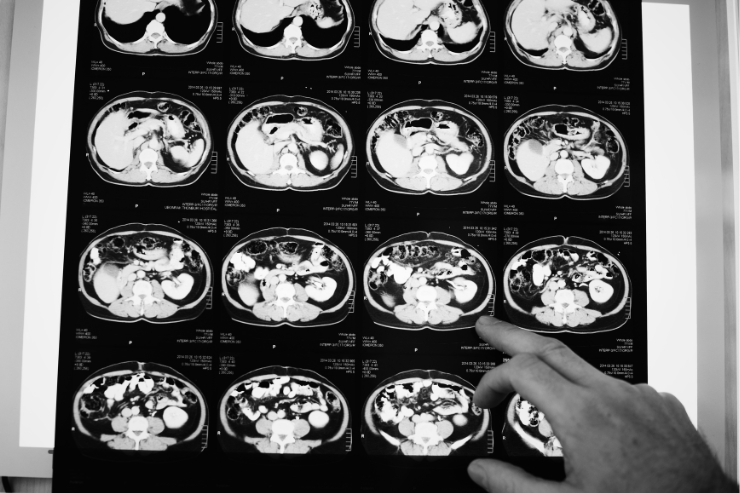
From Raw Images to Insights: The Process of Labeling Medical Data Healthcare AI is reshaping the medical field by providing powerful tools for diagnosis, treatment planning, and patient care. By leveraging machine learning, AI can process complex medical data, uncover patterns, and assist in critical decision-making. However, the accuracy of these AI systems depends heavily on high-quality, annotated data. Medical data comes in many forms—images from diagnostic scans, patient records, and health app data. While these datasets are rich in information, they often lack the structure and labeling needed for training AI models. This is where medical image annotation plays a pivotal role. It provides the precise labels that serve as the foundation for building reliable and accurate AI systems.In this blog, we’ll delve into the process of medical image annotation, the challenges it presents, and why it is so essential. We’ll also guide you on selecting the right annotation tools and partners, showing how this critical step is driving innovation in healthcare AI. What is Medical Image Annotation? Medical image annotation is the process of adding detailed information to medical images, such as MRIs, CT scans, and X-rays, to make them understandable to AI systems. It acts as a bridge, enabling AI models to interpret these images as accurately as a trained medical professional. By marking specific areas, labeling key features, and highlighting subtle patterns, annotators provide the extra information AI needs to analyze these images with accuracy. For example, medical image annotation could involve outlining the edges of a tumor, identifying subtle changes in tissue, or labeling key anatomical structures. These precise annotations are crucial for training AI models to interpret medical data with high accuracy. With these detailed labels, AI can support critical tasks such as diagnosing diseases, planning surgeries, and monitoring treatment progress. What sets medical image annotation apart is the level of precision required, along with the essential role of medical expertise to ensure the accuracy and reliability of the annotations. Type of Annotation In Medical Images Bounding Box Annotation This is one of the simplest and most widely used techniques. A rectangular box is drawn around areas of interest, such as tumors, lesions, or fractures. The bounding box helps AI models localize and identify objects within the image. While this method is effective for detecting large objects, it may not be as precise for irregular shapes, which can lead to less accurate results in some cases. Polygon Annotation For objects with irregular shapes, polygon annotation is used to outline boundaries more accurately. By placing a series of points around the object, annotators can draw polygons that conform to the exact contours of the area of interest. This method is particularly useful for marking regions such as tumors or blood vessels that don’t fit neatly into a box, providing a higher level of precision than bounding boxes. A computer tomography image of brain and skull showing large intracerebral hemorrhage or hemorrhagic stroke. Segmentation A. Semantic Segmentation: In this type of annotation, each pixel in an image is assigned a class label, indicating the type of tissue, organ, or anomaly present. For example, all pixels representing healthy brain tissue might be labeled one color, while pixels corresponding to a tumor would be labeled another. This allows AI systems to understand the full context of the image at a pixel level, which is essential for tasks like diagnosing diseases or detecting subtle abnormalities. B. Instance Segmentation: Unlike semantic segmentation, which groups all objects of the same type together, instance segmentation distinguishes between individual instances of the same object. For example, if there are multiple tumors in a scan, each tumor would be identified as a separate entity. This technique is crucial when there are overlapping or closely located structures that need to be identified individually, such as multiple nodules in a lung scan. Key Point Annotation Key point annotation involves marking specific points of interest within an image, typically anatomical landmarks such as joints, blood vessels, or nodules. These points are often used in AI models to track movement (e.g., in orthopedic imaging) or to identify specific features like the location of a tumor or cyst. Key point annotation is also vital for tasks such as facial recognition or skeletal analysis in radiology. Landmark Annotation Landmark annotation is used to identify and mark specific, fixed points in an image that are crucial for understanding the overall structure or function. These landmarks are usually anatomically significant features, such as the position of a tumor relative to surrounding tissues or specific joints in a musculoskeletal image. Landmark annotation is essential for tasks that require understanding the spatial relationships between different anatomical structures, like preoperative planning or organ segmentation. Process of Medical Image Annotation The process of medical image annotation involves several key steps to ensure the images are accurately labeled and ready for AI training. This process requires a combination of technical expertise and medical knowledge to ensure the highest quality data for AI models. Here’s a breakdown of the main steps involved: Understanding Image Formats Medical images are typically stored in specific formats like DICOM (Digital Imaging and Communications in Medicine) and TIFF (Tagged Image File Format). DICOM is the standard format used in medical imaging, and it includes both the image data and relevant metadata such as patient information, image acquisition details, and machine specifications. TIFF, on the other hand, is often used for storing high-quality images without loss of detail. These images are usually the starting point for the annotation process. 1. Processing DICOM and TIFF Images Before annotating, the images need to be processed to make them suitable for analysis. This may involve converting the raw DICOM or TIFF images into a more manageable format, such as converting 3D scans into slices for easier analysis or enhancing the image quality for clearer visualization of features. This step is crucial because the quality and clarity of the images directly impact the accuracy of the annotations. 2.Choosing the Right Annotation Tool Selecting the appropriate annotation tool
From Raw Images to Insights: The Process of Labeling Medical Data Read Post »